HESCH INSTITUTE
Specialist in Sacroiliac Pain
  • Home
  • About Us/Blog
    • Our Method
    • Qualifications
    • Testimonials>
      • Feedback From Clients
      • Professional Comments
      • Seminar Comments
      • Distance Learning Comments
    • Endorsed Links
    • Jerry's Blog
    • Contact Us
  • Patient Info
    • Local Vegas New Patient Info
    • Out-of-Town New Patient Info
  • Research & Publication
    • Research Supporting Hesch Method
    • Research & Discussion Topics>
      • Hesch Method Basics
      • Manual Therapy>
        • Regional Interdependence
        • Righting Reflex
        • Joint Mechanoreceptors
        • Hypomobility & Hypermobility
        • Pelvis: Cervical Compensation
        • Enhanced Mobilization Scale
      • Professional Library>
        • Cuboid Syndrome>
          • Cuboid Syndrome Overview
          • Cuboid Correspondence
          • Cuboid Photos
          • My (Former) Cuboid Syndrome
          • Cuboid Q & A
        • Coccyx
        • Foot, Ankle, Knee, Hip>
          • Ankle Injury
          • Talus
          • Fibula
          • Peroneal Tendonitis
          • Foot & Ankle Motion
          • Letter: Two Hip Studies
          • Trochanteric Bursitis
        • Inguinal Canal>
          • Inguinal Neuropathy Test
          • Pain After Hernia Surgery
          • Spermatic Cord Mobilization
          • Prolotherapy for Inguinal Pain
        • Low Back Pain>
          • LBP Study
          • LBP Abstract
          • SIJD / LBP
          • SIJ Radiology / LBP
          • LBP Case Study
        • Pregnancy & SIJ>
          • Pregnancy Hyper & Hypomobility
          • Pregnancy Outcome Study
        • Shoulder>
          • Rotator Cuff
          • A/C Joint
        • Sacroiliac & Pelvis>
          • SIJ Dysfunction: Most Common Pattern
          • Ilial Anterior Rotation Hypermobility
          • SIJ Spring Test Article
          • Problems with Traditional SI Evaluation
          • Sacrum: Key Concepts
          • Spring Test
          • Downslip Ilium
          • SIJ in Low Back Pain
          • SIJ Radiology
          • McConnell Taping
          • Side Glide Dyfunction
          • Sacral Rotation
        • SIJ Miscellaneous>
          • Letters to the Editor: SIJ
          • SIJ Blog Comment
          • SIJ Dialogue
        • Symphysis Pubis>
          • Symphysis Pubis
        • Thoracic Spine & Ribcage>
          • Ribs & Thoracic Segments
          • Manual Therapy Sternum
          • Cervical Pain Thoracic Manipulation
        • TMJ & Cervical Spine>
          • TMJ / Upper Cervical
          • Righting Reflex
        • Other Topics>
          • Hesch Method Course Evaluation
          • Interdisciplinary Study
          • Three Dimension Pain Drawing
          • Arachnoiditis
          • Letters To The Editor
          • Blog Postings
      • Video Library>
        • Upslip vs. Downslip
        • Lower Pelvis & Coccyx
        • Pubic Symphysis
        • Sacrum
        • Complex Pelvic Dysfunction
        • Foot
        • Shoulder
        • Knee
        • Regional Interdependence Videos
        • Precourse Lab Practice
        • Post-course Videos
    • Articles & Information
    • Hesch Publication
  • Education
    • Our Mission: Education
    • Seminars>
      • Seminar Registration
    • Distance Learning>
      • Distance Learning Registration
      • DL Video Files
      • DL Advanced Video Files
      • Dynamic Body Torsion Chapter
    • Expos & Events
    • 3D Pain Drawing
  • Store
    • Instructional Videos
    • Products

Cuboid Photos

Picture
Lateral view of right cuboid. Arrow Points to the right cuboid, lookin from the outside of the right foot..
Picture
Top view of left cuboid.
Picture
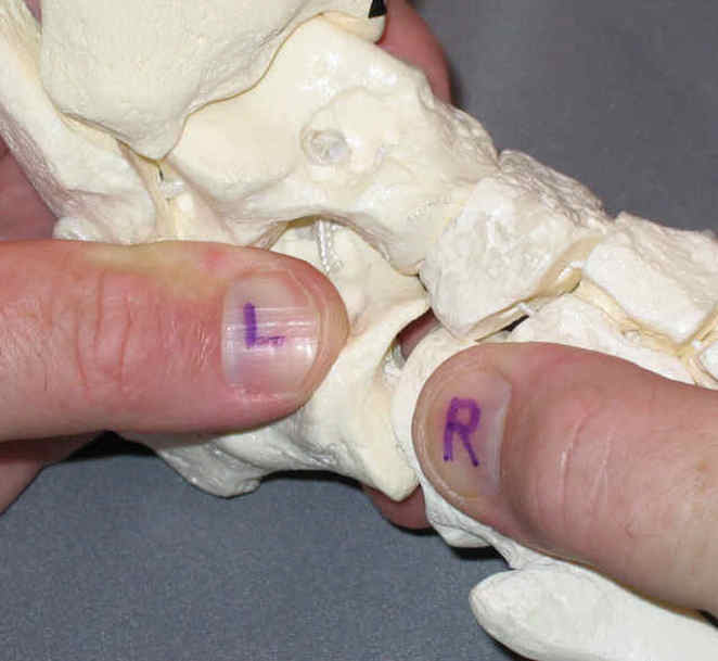
Outside view of left foot. Left thumb is on the calcaneus (heel) the largest bone in the foot. right thumb is on the cuboid. I am distractiin the joint and you can see the very curved joint surface. the "take home message" is that the calcaneus is much larger and has a powerful influence on the cuboid. It is very important to evaluate mobility of the calcaneus and restore normal mobility when able, as opposed to just restoring mobility of the cuboid, in presentations of cuboid syndrome. The goal here is prevention of recurrence, not just relief of acute symptoms.    
Picture
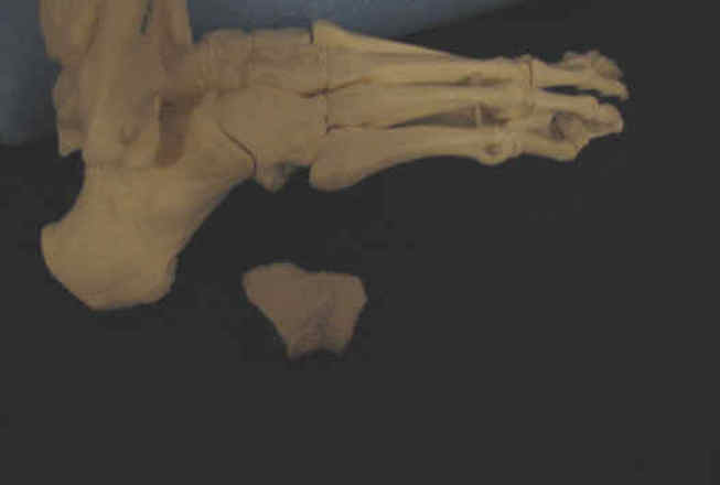
Lateral view of right foot with intact cuboid and solitary cuboid below.
Picture
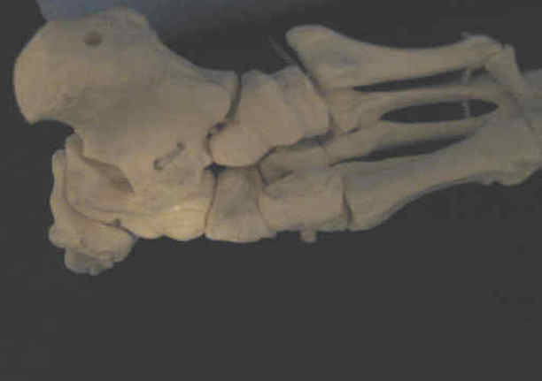
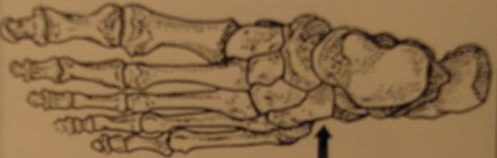
View from bottom of foot. Note the curved surface where the calcaneus (large bone top left) joins the cuboid, whereas other bones that connect to the cuboid have a fairly flat (planar) shape.