|
One of my mentors Phillip Greenman, DO passed away on February 7, 2013. Below
is a quote from the sacral torsion chapter I wrote acknowledging his influence on my work. He has influenced many. "I wish to express a debt of gratitude to the osteopathic profession, and in particular to Philip Greenman, DO, who greatly influenced my development as a hands-on clinician. I hope my reinterpretation honors your vision and your body of work." 
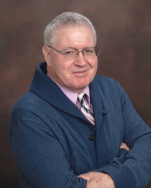
Philip E. Greenman, D.O. Philip E. Greenman, D.O., passed away on February 5, 2013 in Tucson, AZ due to complications of pneumonia. Dr. Greenman was born on February 25, 1928 in Deposit, NY, the only son of Joseph and Thelma Greenman, and was a 1952 graduate of the Philadelphia College of Osteopathic Medicine. He was in private practice in Buffalo, New York for almost twenty years before accepting a position at Michigan State University in East Lansing, MI in 1972, where he served as Professor and Associate Dean of the College of Osteopathic Medicine before retiring to Tucson in 2004. Dr. Greenman authored a noted medical textbook and was internationally known for his work and research in the field of manual medicine. He is survived and fondly remembered by his wife of 63 years, Patricia Bingham Greenman, his sons John and Jeffrey, daughters-in-law Laura and Janet, and grandchildren Elizabeth, Alexander, Emily, Matthew and Andrew. A memorial service will take place at Grace/St. Paul's Episcopal Church in Tucson at a later date. Memorial gifts would be welcomed for the Philip E. Greenman Endowed Residency (AS040) by sending a check payable to "Michigan State University" to MSU College of Osteopathic Medicine, 965 Fee Road, Room A310, East Lansing, MI 48824. THEORY REPORT
REAPPRAISAL OF SOME FUNDAMENTAL MANUAL THERAPY CONCEPTS Hesch J, Hesch Institute, Henderson, NV Corresponding author: [email protected] Every profession benefits from periodic appraisal of their foundational concepts. It is encouraging to note fundamental reappraisals of basic tenets such as recent works on evaluating concave-convex rule, and potential belief such as grade V manipulation being superior to grades I-IV mobilization. Other foundational beliefs and scales may be timely for reappraisal. Qualifiers may enhance the traditional joint movement scale. For example Grade I, II hypomobility can include a fixed, immutable hypo and hypermobility such as in the former; from genetics, age, disease, scar tissue, and for the latter etc. Treatable hypomobility may also be due to proximal or distal reflex or biomechanical faults, muscle guarding, etc. Similar rationale may apply to grades IV and V hypermobility. The mobilization scale could include a separate grade for constant force maintained at end of available range for an extended period of time, AKA viscoelastic creep. Non-synovial joints with dense connective tissue such as the SIJ, symphysis pubis, or joints such as the subtalar with an interosseous ligament, or joints with long-lasting restriction may benefit from creep mobilization. Another concept is that of end feel. A recent case presented with a bone on bone end feel of both hips with ten degrees of internal rotation. Initially reluctant, the author ultimately treated it with a gain to 45 degrees within two sessions. A separate topic for exploration would be the limits imposed by language and by categorization, and the value of clarifying terminologies within manual therapy, medicine and the overall clinician-client interaction. Several members of a facebook sacroiliac joint group have informed me that SI-Bone has made their SIJ fusion device unavailalbe to a highly marketed SIJ fusion group. Sometime last year they removed that group from their website where they had a prominent presence. I submit that a very small part of failed back pain population may benefit from SIJ stabilization but that for many it is not adequately determined and worse: we/they especially physical therapists who promote SIJ fusions DO NOT KNOW WHEN NOT TO RECOMMEND FUSION. I am in touch with some of the horrific failures, and hope for a much improved, and much more thorough algotithm...pray hard and pray fast and take action.
APTA President responds to Dr Oz producer regarding "cutting-edge treatment" for low back pain2/7/2013
Dr. Paul Rockar, President of the APTA, issued a press release today regarding
the recent segment in which a Physical Therapist discussed cutting edge solutions for LBP on the Dr. Oz show. The statement is direct and I for one, applaud this effort. See it for yourself: February 5, 2013 Dear Producer: APTA takes exception to the portrayal of ultrasound, Tiger Balm patches, and bumpy balls as “cutting-edge physical therapy treatments” for back pain in your recent segment “Cutting-Edge Solutions for Back Pain.” While modalities may be used by physical therapists as part of an overall treatment plan, the focus of physical therapy treatment for back pain is on evidence-based exercises to improve strength and flexibility, manual therapy to improve the mobility of joints and soft tissues, and patient education on ways to enhance recovery, prevent and relieve pain, and avoid recurrence. These avenues of care offer long-term solutions rather than temporary, intermittent relief. Pleased to announce this was accepted for the American Academy of Orthopedic Manual Therapy 2013 conference. As a platform paper or poster; TBD
Case Study Report THREE CASES: PREDICTIVE VALUE OF PASSIVE PELVIC MOTION TESTING IN EARLY INFLAMMATORY SPONDYLARTHROPATHY AND IN SACROILIAC FUSIONS FROM H1N1 VIRUS INFECTION AND FROM HARDWARE INVASION VALIDATED WITH CT SCAN Hesch J Hesch Institute, Henderson, NV, USA Corresponding author: [email protected] Background and Purpose: Three cases with loss of passive pelvic macromotion (PM) and sacroiliac joint (SIJ) micromotion (SIJM) are presented. Case one (C1) had an H1N1 virus infection with pathological left SIJ fusion. Case 2 (C2) presented with an extensive thoracic and lumbosacral fusion who requested consultation, anticipated a SIJ fusion. Case three (C3) was an athlete treated because of significant and lasting drop in performance. Description: The purpose of this case series is to compare passive SIJM and PM loss with objective imaging (CT scan) and explore diagnostic utility of SIJM and PM in a case of early, not-yet-diagnosed inflammatory spondylarthropathy (IS). Outcomes: Unilateral loss of PM and SIJM in C1 and C2 correlated positively with unilateral SIJ fusion per CT scan. Normal joint space at S1 and S2 and absence of joint space at S3 from hardware invasion was observed in C2. Inability to gain PM, SIJM in C3 aroused suspicion of SIJ pathology and provoked rheumatologic referral. Discussion and Conclusion: The PM and SIJM tests correlate with fused SIJ’s, have utility for a subset of subjects who contemplate SIJ fusions, and for early detection of IS in which early SIJ fibrotic changes may escape early detection, delaying definitive diagnosis up to 10 years from initial onset. OBSTETRIC PUBIC SYMPHYSEAL DIASTASIS: IMAGING SUPPORT OF A NOVEL BIOMECHANICAL MODEL Hesch, J Hesch Institute, Henderson NV, USA Introduction In pregnancy pelvic joint and pelvic outlet widening occurs in response to ligamentous softening, fetal growth, and parturition. Obstetric pubic symphyseal diastasis (OPSD) is widening of the symphysis at mid-joint of 10mm or more. Progressive widening can provoke severe pain and functional impairment. Most recover with conservative care; surgical stabilization is typical for 25mm or greater. Chronic painful pubic dysfunction due to OPSD is a poorly understood disabilitywth chronic widening of less than 10mm, pain and functional limitations. MRI and CT images undergird novel biomechanical interpretation, suggesting treatment modification. Purpose To present a novel hypothesis that OPSD involves a distinctly different biomechanical trajectory rather than simply a traumatic escalation of peripartum pelvic mechanics. Materials and Methods Articles gathered from www.PubMed.org and general web searches using key words symphyseal diastasis, pubic joint instability, obstetric instability, and pubic instability, covering 1997-2012. MRI and CT images were evaluated in cases of acute OPSD with gapping of anterior-superior SI joint (SIJ). Results Several images demonstrated SIJ gapping with a peculiar previously unreported, inferior-posterior retroarticular approximation; provoking further inquiry. Testing with ligamented pelvis and flexible anatomical models was performed to simulate obstetric joint and outlet widening. This included sacral nutation, medial infolding of the ilia, and spreading of the ischial bones, lower pubic and SIJ, as described in the literature. A distinct compressive end-point was encountered in the superior-anterior SIJ and superior pubic joint, where no further expansi on could occur. Diastasis was introduced by cutting and separating the joint. Relevance Replicating the gapping of the superior SIJ, congruent with the OPSD x-ray, MRI and CT images, actually reduced pelvic outlet dimension. Only by moving the pubes and ilia in a cam-like manner, coupled with posteromedial glide at the SIJ’s, primarily in the transverse plane, was pelvic outlet maximized. No other trajectory was able to enhance outlet dimension to this degree. Conclusions Imaging and mechanical testing support the hypothesis of novel biomechanics with OPSD. Research using this hypothesis may identify optimal intervention for acute and chronic cases. Discussion The unique properties of the axial ligament; size, location and decreased elastin, suggest that in the presence of OPSD it may function as a mechanical stop during parturition, maintaining optimal pelvic outlet dimension. In chronic cases, muscular and ligamentous recoil may be absent or insufficient, preventing form-closure. The chronic pubic dysfunction and OPSD populations are especially in need of improved care. In vivo biomechanical research during parturition is improbable. Computer modeling and pre/post intervention imaging and functional metrics seem reasonable. Implications A diagnostic tool may already exist in the form of CT or MRI images in the medical record. Passive external forces directed at reducing the unique cam-like transverse plane movement of the ilia prior to or during application of a pelvic binder at the trochanters, an orthopedic standard of care may have merit. Keywords: obstetric pubic symphyseal diastasis, SIJ, axial ligament, pelvic binder. http://erikdalton.com/low-back-pain-foot-posture/
It is a nice article though the description of the cuboid mechanics is not congruent with what I find clinically. Also, the description of the definition for pronation and supination change from non-weight bearing as described to weight bearing due to the forces of the ground resisitng abduction and adduction; which for the most part are confusing terms indeed. A deeper exploration should explain the difference between forefoot and rearfoot pronation/supination because some feet have for example rearfoot supination and forefoot pronation, especially in weight bearing and the perspective of the clinician becomes very important in communicating terminology, using terms such as compensated and uncompensated. I am proud to be one of the clinicians who influenced some of Erik's work on the SIJ/pelvis through my home study course. Its OK Jerry you have worked hard and so much of your work is gratis, OK to share your pride SACROILIAC FUSION WITH "ALIGNMENT BY A PHYSICAL THERAPIST" IS A BELIEF SYSTEM, NOT SCIENCE1/23/2013
“Let me first differentiate between belief systems and science.
A belief system is a body of concepts, ideals or narratives that through written or oral tradition and historical precedent have become an integral part of a culture. Verification is dependent on faith in its tenets: Ergo—subjective. A belief system need not concern itself with objective reality. This contrasts with science in which theories are subject to objective evaluation by repeated experiment and measurement. Science assumes a priori the existence of a measurable objective reality. Indeed, science is the delineation of this reality. The precision of the replication of experimental results and the accuracy of theory in making predictions, even when both observers and theorists may initially have a wide variety of theoretical considerations, is the affirmation of the scientific approach to the comprehension of the cosmos.” * SIJD with sub-clinical (cannot be measured objectively such as with x-rays, MRI’s CT scans, etc., qualifies as a belief-system diagnosis. Severe pelvic instability is science. It is measured with x-ray, CT scan, MRI and objective measures such as degrees of separation, whether at the symphysis pubis joint (primary instability with intact SIJ) or symphysis pubis instability with subsequent SIj instability. See Tile classification system. For any and all who travel to see SIJ “experts” to see if they need a SIJ fusion with adjustment (a completely subjective thing), you are entering a world that is objectively named: A BELIEF SYSTEM. Jerry Hesch, MHS PT *
This is a very kind and thoughtful feedback from a recent client. The feedback demonstrates the value of a whole-body articular evaluation and treatment. I believe also that the work paved the way for clarifying that which was amenable to hands-on care, and that which was not, rather than being a vague interlinked unclear pain generator. I am referencing the inguinal pain. I will write back and share some other thoughts to consider re the inguinal pain as part of her medical work up. I am very grateful for this kind feedback.
Dear Jerry, I just want to thank you again for helping me out with my back. I feel like I am getting my life back. Medical Doctors, Chiropractors, Acupuncture, Physical Therapy, Physiatrists and Pain Specialists were not able to accomplish in nearly a year and a half (and more than 50 office visits) what you accomplished in twenty to thirty minutes during my first visit. I am still in awe of the fact that I am no longer in constant pain. It has been nearly two months since you treated me and I am continuing to improve in many respects. I am still dealing with some nerve irritation when I sit but I have no back pain at all from sitting. I can walk as long as I like. I am able to do most chores around the house without paying the price afterwards. I am even back to work and teaching a class online this quarter for the university since I can sit with no back pain. I can also still move and turn my mid back at the thoracic lumbar junction where it was frozen before. I am spreading the word about the great work you do. I have shared with my local physical therapist, pelvic floor therapist and the pain doctor I was seeing in Seattle. I have not had to see any of them since I saw you in November. I also shared information with my cousin, (the OB/GYN from Bend, Oregon who also suffers from terrible back pain in the lumbar and SI region) whom I mentioned when I saw you in November. She is very impressed that I am still feeling great nearly two months later. I believe she too will be contacting you if she already hasn't. My daughter, is doing well. She occasionally has some knee pain but not with the same frequency as she had before she saw you in November. Thank you so very much Jerry. I will keep you posted and I will continue to share with people who have treated me and people who could use your help, the great work that you are doing and giving people their lives back. Sincerely, Chantelle Hildreth Spine:
POST ACCEPTANCE, 15 January 2013 doi: 10.1097/BRS.0b013e318286b7dd Clinical Case Series: PDF Only Do L5 and S1 nerve root compressions produce radicular pain in a dermatomal pattern?Taylor, CS BSc, MBBS; Coxon, A PhD, MBCS, MICR; Watson, P BSc; Greenough, CG MD, MChir, FRCSPublished Ahead-of-Print Abstract Structured Abstract: Study Design. Observational case series. Objective. To compare the pattern of distribution of radicular pain with published dermatome charts. Summary of Background Data. Dermatomal charts vary, and previous studies have demonstrated significant individual subject variation. Methods. Patients with radiologically and surgically proven nerve root compression caused by prolapsed intervertebral disc completed computerised diagrams of the distribution of pain and pins and needles. 98 patients had L5 compressions and 83 had S1 compressions. Results. The distribution of pain and pins and needles did not correspond well with dermatomal patterns. Of those patients with L5 NRC, only 22 (22.4%) recorded any hits on the L5 dermatome on the front, and only 60 (61.2%) on the back with only 13 (13.3%) on both. Only 1 (1.0%) patient placed >50% of their hits within the L5 dermatome. Of those patients with S1 NRC, only 3 (3.6%) recorded any hits on the S1 dermatome on the front, and only 64 (77.1%) on the back with only 15 (18.1%) on both. No patients placed >50% of their hits within the S1 dermatome. Regarding pins and needles, 27 (29.7%) of L5 patients recorded hits on the front alone, 27 (29.7%) on the back alone and 14 (15.4%) on both. 19 (20.9%) recorded >50% of hits within the L5 dermatome. 3 (3.6%) S1 patients recorded hits on the front alone, 44 (53.0%) on the back alone and 18 (21.7%) on both. 12 (14.5%) recorded >50% of hits within the S1 dermatome. Conclusion. Patient report is an unreliable method of identifying the anatomical source of pain or paraesthesia caused by nerve root compression. |
Dr. Jerry Hesch, DPT, MHS, PTMarried with 4 grown kids. Earned my Doctorate at A.T. Still University in Tempe, AZ, MHS at the University of Indianapolis and my BS PT at University of New Mexico. I enjoy working with my hands and particularly making glass objet d'art. Powered by Calendar Labs Archives
August 2016
Categories
All
|
RSS Feed